Tuesday, June 16, 2015

A mutually dependent Relationship: Diabetes & Obesity


              A mutually dependent Relationship: Diabetes & Obesity
Obesity and Diabetes are pandemics that are infiltrating into the world population at very alarming rates. Basically, as per the indication from the Centre of Disease Control, a big portion of the world population is living with obesity. For instance, statistical figures reveal that, approximately seven percent of the American population is having obesity, accounting for close to 59 million Americans something that has become on the biggest challenge to community vitality. Statistics for the last decade reveal that;

  • The rate of obesity has increased to 56 percent while diabetes has increased by 5 percent
  • The future generation is likely to be immensely affected by diabetes
Basically, the risk factors for both diabetes and obesity are linked to age, gender, history of the family and race. However, it is important to understand that the modern things we do now contribute immensely to the expansion if the two sicknesses. The example of life style issues includes little or no physical exercise and the like of food stuffs that have high fat contents.


These foods are known to cause obesity, but the question remains; do the same foods cause diabetes?
What is the connection between diabetes and obesity?
As it has always been the case, is more often than not, most of the patients who are diagnosed with diabetes as well have obesity. As a result of this observation, we can comfortably tell that there must be a link between obesity and diabetes. So as to prevent this disease in future times, it is critical that one should get informed about the major causative factors.

When you are fat, the body becomes very heavy. As a result, much of your weight will be exerted on the body in many ways. One of the ways can be the inability of the body to keep blood glucose level at optimal levels. As a matter of fact, a lot of body mass resists against insulin thus the body does not utilize insulin well. Basically, the form in which absorbed glucose is reserved is fat. The more glucose is converted into fat results in addition of body mass, thus obesity. It is important that, when on insulin intake, one should avoid taking a lot of foods to prevent diabetes,http://www.joslin.org/info/why_did_i_gain_weight_when_i_started_taking_insulin.html.
Preventing obesity will prevent diabetes too
By virtue of the causes diabetes and obesity, it is apparent that observing some basic lifestyle issues will help reduce or treat the two diseases. These are very steps whose impacts are far reaching. You need to do some physical exercise very day as this will help extra fats in the body, eat a balanced diet; that is to mean that the diets should have too much of fats and in some cases, use vitamin supplements tailored to diabetic patients.

In some cases, the patient could be obese. As such , it is highly recommended that one looses a bit of their weight which will in turn lower the risks of diabetes. Weight loss can be difficult but the following components of diet can quicken up the process; low levels of carbohydrates, high fiber content coupled with a workout plan.

                                                  References


Sunday, June 14, 2015

Research Chimps Now Considered Endangered Species

Two days ago on June 12, 2015, the United States government is now considering chimpanzees used for research as an endangered species through the Endangered Species Act. This means that researchers cannot use chimps for medical experiments as freely as they have before. Before this adjustment, “The United States is the only major country that still funds invasive chimpanzee research,” according to Nature Publishing Group. The only way that chimpanzees can be used now is if the person has a permit from the U.S. Fish and Wildlife Service. Many regulations for research using chimp have been made such as “work enhances the survival of the species and benefits chimps in the wild”, if the chimp is being used for human purposes it must be crucial to a human disease, the researcher must somehow support chimpanzee preservation, and follow standard research protocol such as applying for project approval and undergoing inspections (AAAS).
This change has affected many different people in positive and negative ways. For animal rights groups and specific chimpanzee groups like the Jane Goodall Institute, it is a huge success for something they have been working towards for many years. The director of the U.S. Fish and Wildlife Service was quoted at the press conference announcing this decision, “This decision will help us ensure that the world we pass along to our children and grandchildren will be filled with chimpanzees. We believe this action will ensure that activities affecting all chimpanzees will contribute to the survival of chimpanzees in the wild,” (AAAS). The chimps being released from laboratories will be sent to available sanctuaries since there is no possible way of letting them go into the wild.
For laboratories using chimpanzees for research, this modification will somewhat affect their work. If they wish to continue using chimps they have to apply for strict permits and be subject to more inspections, as well as supply some type of funding to protect wild chimpanzees. An anatomist at Stony Brook University, Susan Larson, was quoted, “This is going to make it increasingly difficult to get these projects off the ground,” (AAAS).  But Larson is in the minority because many other institutions who have done chimpanzee research in the past are okay with this change in chimp status. There is a lot of recent new technology, such as in vitro testing using cells grown in a petri dish, which can replace the use of animal testing. If chimp research is absolutely vital to the development and the betterment of human life then they can apply for a permit. Many research facilities have actually already gotten rid of animal testing due to pressure from animal rights groups and popular public opinion.

Although animal testing has led to many scientific discoveries and advancements for humanity, we are now at a point in time that scientists can agree that it is not entirely necessary. This change from research chimpanzees to endangered animals will not have a large effect on human life and is a positive step for animal rights. 

http://animal-testing.procon.org/#pro_con

Saturday, June 13, 2015

Ricin


I recently watched a Youtube video about natural poisons. One of poisons that they talked about was Ricin. I had heard of this poison before in a TV show or movie before so I decided to read more about it. This is what I found.
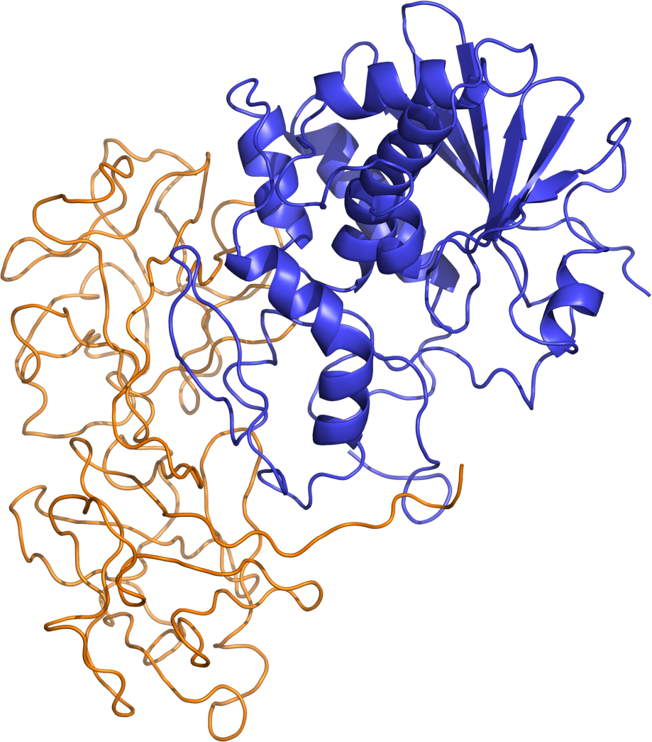
Ricin structure.

Ricin is known as a highly toxic lectin which is naturally occurring. lectins are one of many types of proteins that can bind to a cell's membrane. Ricin is produced by in the seeds of the castor plant. According to the CDC  "the LD50 by intravenous injection is about 5 µg/kg, whereas it is 20 mg/kg by intragasteric route.23,24 The ricin aerosol LD50 for NHP is estimated to be 10-15 µg/kg.17 The human lethal dose has not been established rigorously, but may be as low as 1-5 mg of ricin by injection 25 or by the aerosol route" This shows just how little Ricin is needed to kill a human.

Just some quick backstory on what median lethal dose is means. Median lethal dose its a means to measure how lethal a toxin is. It means that its dose required to kill half the members of a tested population after a specified test duration. LD50 is simply and abbreviation meaning lethal dose 50%. A lower LD50 number means more toxicity. This test was first done in 1927 by J.W. Trevan. The main way to find the LD50 is to simply test the substance on lab rats. The LD50 test can be some what unreliable as the variation is so great between different species' immune system and physiology.

Ricin has been around for some time. according to Globalsecruity.org "During World War I, the United States explored ricin as a potential offensive biological weapons toxin at the American University Experimental Station. Two methods of weaponizing ricin were explored: bullets and shrapnel coated with ricin toxins or a 'dust cloud' of toxin toxins inhaled into the lungs." Recently some terrorist groups have experimented with using ricin as a weapon and caused several incidents of the poisons being mailed to U.S. politicians. For example, A letter containing ricin was allegedly sent  to American President Barack Obama at the same time.

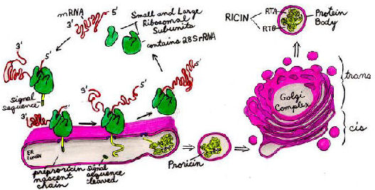
Ricin is extremely toxic if inhaled, injected, or ingested. It kills by slowing or stopping protein synthesis. This means that ricin prevents cells from assembling amino acids into proteins. A Cornell university resource goes much more in-depth about this. According to the Cornell university college of agriculture and life sciences "Ricin inhibits protein sythesis by specifically and irreversibly inactivating eukaryotic ribosomes." It goes on to say "These "ribosome-inactivating proteins" (RIPs) are typically N-glycosylated, 30 kDa monomers (Type 1 RIPs). However, in order to bind to the cell surface galactosides and enter the cytosol to reach ribosomes, they require a second monomer, a galactose-binding, 30 kDa lectin. The monomers are joined by a disulfide bridge to form the toxic heterodimers (Type 2 RIPs)." Here is a diagram showing the life cycle of ricin as it inter acts with the cells.

Sources:

Helicobacter Pylori, a Little Bacteria with Big Consequences

From the sixth grade until the eighth grade I had debilitating pain in my abdomen roughly thirty minutes to an hour after each meal that I ate. In addition to that I was always bloated, frequently nauseous after I started to eat, and no matter how much I ate I would lose weight. My doctor was baffled at what it could be and I endured months of tests--mostly blood work--and doctor’s appointments before he finally figured it out: my stomach was riddled with the bacteria Helicobacter Pylori (H. pylori).
About half of the world's population is actually infected with this bacteria, but most people do not have any symptoms or have any problems linked with H. pylori. In addition to the symptoms that I experienced, one could possibly encounter a loss of appetite or frequent burping.  The complications of H. pylori are far worse than the pain in the abdomen (and it is

not easy pain). H. pylori can lead to an inflammation of the lining of the stomach due to irritation, ulcers, or stomach cancer. Roughly one in ten people with H. pylori develop an ulcer.
When someone comes into the doctor's office with the symptoms of H. pylori, there are several ways that they can be tested for the bacteria. I was diagnosed by a blood test, which is the least accurate way of determining active H. pylori. A blood test simply tests whether or not there are antibodies for H. pylori, which would appear to be positive even if that strand of H. pylori was not harmful or the body had already defeated it.  Another option is testing the breath after taking medication that would give off carbon if in the presence of H. pylori. Certain medicines in your system can affect the validity of the breath test, though. Fecal matter can be tested for antigens that would lead the doctor to the conclusion that H. pylori is present, but the same medicines can affect this test. Lastly, the doctor can sedate the patient and perform an endoscopy exam which involves putting a long tube down the patient's throat with a camera on the end to view the inside of the stomach and to take a biopsy of the tissue to test for H. pylori directly. However, due the invasive nature of this test, it is typically reserved for diagnosing ulcers associated with H. pylori or in a last effort to definitively rule out other conditions. 
Once diagnosed, the treatment for H. pylori is fairly simple. The doctor will typically prescribe two types of antibiotics because of the resilience of the bacteria and its ability to develop a resistance to the antibiotic. On top of that, doctors give medication designed to block acid in the stomach in order to start the healing process of the stomach lining. There are several options for acid blockers such as proton pump inhibitors (ex: Prevacid), histamine blockers (ex: Zantax), or bismuth subsalicylate (ex: Pepto-Bismol). With the resilience of this bacteria, it is also important that the patient is tested at least a month after treatment has ended to see if another round of antibiotics are necessary.  
All of the information dealing with the treatment of ulcers caused by H. pylori can be attributed to Dr. Barry Marshall. In the 1980s, it was thought that ulcers were caused by stress but Marshall knew that was not true—he knew that they were caused by H. pylori. After seeing patient after patient die from severe ulcers that were not caused by stress, he knew that he had to do something about it. The bacteria would not grow in lab mice to be tested, so he infected himself with a very bad strain of H. pylori to see if it would give him an ulcer and then if he could treat it. It is because of this man’s devotion to the health of his patients and this courageous experiment that H. pylori has become severely less deadly and highly treatable.


Sources:


Neonatal Adrenoleukodystrophy

I decided to post about a very rare genetic disorder known as neonatal adrenoleukodystrophy. Most people have never heard of this rare disorder. This is genetic disease that I am very familiar with because my cousin was diagnosed with neonatal adrenoleukodystrophy at the age of 6 months and sadly died at the age of three. By all appearances Andy was a normal newborn but it wasn’t long before my aunt and uncle noticed that something was wrong. Initially the pediatricians told my aunt and uncle that everything was fine and not to worry. Andy did not respond to visual cues or sound. Very soon after birth it was determined that Andy was deaf and blind. He was missing all of the milestones children should hit by the age of 6 months. Andy could barely hold his head up and could not sit up or role over on his own. It was a mystery to my family until he went to a specialist and it was determined that he had a very rare condition called neonatal adrenoleukodystrophy. This genetic disorder affects about 1 in every 50,000 births. This disorder is characterized by the breakdown or loss of the myelin sheath surrounding the nerve cells in the brain which causes dysfunction of the adrenal glands. In class we learned that the myelin sheath is responsible for the transmission of the electrical impulses along the axon. The breakdown of the myelin sheath affects one’s ability to transmit information and this explains why my cousin was unable to see, hear, roll over or even sit up.  Unfortunately, this particular form of leukodystrophy is fatal and there is currently no cure. For this reason my family has been very involved with promoting awareness and supporting fund raisers. Because this disorder affects the nerves and adrenal glands this is a disease that will get progressively worse. This specific form of leukodystrophy is caused by a mutation in the PEX5 gene. For an individual to have this disorder they must have two copies of the mutated PEX5 gene. Both my aunt and uncle were carriers of this mutated PEX5 gene. My aunt and Uncle gave birth to another son several years later and were very lucky not to have passed along this disorder to their second son. Because they were both carries of the recessive mutated gene PEX5 they have a 50 % chance of having a child with the disorder. I found the topic of this blog post interesting not only because of my personal connection but also because of how this disorder relates to multiple topics leaned about and discussed throughout this class. During this class we discussed at length the transmission of recessive alleles and the pedigree analysis and genetic counseling used.  We also discussed the function of the myelin sheath on the neurons axon and what this is responsible for. Currently there is research being conducted using stem cell transplantation using umbilical cord cells and bone marrow cell. Unfortunately these treatments can only slow this degenerative process of the myelin sheath deterioration. The most effective way to prevent this disorder is understanding genetic testing for the recessive alleles.

Work Cited:

Say NO to GMOs

GMO, which stands for genetically modified organisms, are being found in food we eat more so now than in the past. What exactly is GMOs? GMOs are genetically engineered organisms that have been changed and manipulated in laboratories. The reason why GMOs are so harmful to the human body and more people are starting to become GMO free is because these labs are directly spraying chemicals onto the food while it is being processed. GMOs have been added into food without being identified. What this means is, food that we have been eating has been processed with these chemicals and we have been ingesting them every time we eat. Just because a package of food says “organic” does not mean that it is GMO free, either. If something is organic, either no chemicals were used, or the chemicals that were used were “natural.” There is a website called the “non-GMO project” that gives complete lists of GMO free, and verified, foods and different ways to avoid GMOs. This website gives good insight to those who are wanting to learn more about GMOs and what they really are.
              I have recently started to eat GMO free foods. In all honesty, I feel so much better and I do not get as tired as easily as I did before. There are many documentaries on Netflix about food and GMOs which are extremely interesting. One show that I have watched is called, “Food Inc.,” and they talk heavily about food in grocery stores. Most of the foods, especially tomatoes, when they are sold out of season, they are genetically engineered, all the way down to the color. Knowing this about the foods that are being sold, and being allowed to be sold, is a little frightening to me. According to the non-GMO project website, in The United States alone, over 80% of our food is processed with GMOs.
              Since science is always changing, and will continue to always change, all we can do, as consumers, is make sure we are putting the best foods into our bodies. One way I am starting to do this is by composting my own food waste. In the city that I live in, we actually have compost bins for everyone to use. This is helping to grow organic foods, with no chemicals. The gardens where these foods are being grown are available for anyone to come and pick from the harvest. I think this is a very important step in the right direction. The next important step is to continue to spread the word about the compost bins, and teaching kids and families about eating organically. According to another article, listed below, Holland has been testing GMOs on mice. When they studied these mice, they noticed drastic behavior changes between the mice who have been given the GMOs and the mice who have not. Even though this has only been tested on mice, I hope more tests are done to see if GMOs have this type of effect on students and people with behavior and social issues.

Also attached is a video on GMOs from Bill Nye.

Non-GMO Project


Mice on GMOs Article


Wednesday, June 10, 2015

Lack of Sleep May Lead to Alzheimer's Disease

Not getting enough sleep may be a leading cause of Alzheimer’s disease. Some new studies have shown that a lack of quality sleep may lead to a buildup of a protein that is shown to cause Alzheimer’s.  “Scientists at the University of California, Berkeley, have found compelling evidence that poor sleep -- particularly a deficit of the deep, restorative slumber -- is a channel through which the beta-amyloid protein, believed to trigger Alzheimer's disease, attacks the brain's long-term memory” (University of California – Berkeley, 2015).  They believe that the lack of restorative sleep allows the beta-amyloid protein to build up, causing a weakening of the memory.

Beta-amyloid starts out as a single molecule but these molecules tend to bunch up together.
As they bunch up they form a plaque that can be seen in Alzheimer’s patients. A study at Stanford University School of Medicine showed that “for the first time that in this clustered form, beta-amyloid can bind strongly to a receptor on nerve cells, setting in motion an intercellular process that erodes their synapses with other nerve cells” (Stanford Medicine, 2013). We learned in class that the synapses are how the cells send and receive electric charges in the nervous system. If these synapses get blocked the cells cannot function properly and can lead to the cells death. As more and more of the cells die this leads to severe memory loss which is the hallmark for Alzheimer’s.

It has been shown that there is a buildup of beta-amyloid in people who had Alzheimer’s and those who had a sleeping disorder. “ Moreover, a 2013 University of Rochester study found that the brain cells of mice would shrink during non-rapid-eye-movement (non-REM) sleep to make space for cerebrospinal fluids to wash out toxic metabolites such as beta-amyloid” (University of California – Berkeley, 2015).  This helps to show that sleep can help to get rid of some of the buildup of this toxic protein. They are still trying to figure out whether a buildup of the protein causes poor sleep or if poor sleep causes a buildup. They are still doing research on which is the leading factor.

To see how sleep affects the memory and the buildup of the beta-amyloid in people researchers at Berkley did a study where they had people memorize a list of words and then go to sleep to see how much they remembered the next day. Using brain imaging and other diagnostic tools they were able to show that those who got a good night sleep had a lower level of beta-amyloid and were able to remember more the next day than those who had the poorest quality of sleep.  This shows that the quality of sleep we get can affect the amount of beta-amyloid.
Heavy deposits of the toxic protein, beta-amyloid, shown in red in the brain on the right, are linked to poor sleep and may be paving the way for Alzheimer’s disease. A brain benefiting from deep sleep brain waves and an absence of beta-amyloid is shown on the left.
Credit: Photo courtesy of Bryce Mander and Matthew Walker

If sleep is the leading cause of Alzheimer’s this may be considered a good thing. Getting more sleep is treatable. There are many ways that people can increase their amounts of sleep including exercise, therapy, and maybe even electrical stimulation that amplify the brain waves during sleep. Just getting enough sleep could maybe decrease your likelihood of getting Alzheimer’s or decrease the effects of the disease. More studies are going to be done to solidify the link between sleep and Alzheimer’s. 
Works Cited:
University of California - Berkeley. "Poor sleep linked to toxic buildup of Alzheimer's protein, memory loss." ScienceDaily. ScienceDaily, 1 June 2015. <www.sciencedaily.com/releases/2015/06/150601122442.htm>.

Stanford Medicine. "Scientists Reveal How Beta-amyloid May Cause Alzheimer's." News Center. Stanford Medicine, n.d. Web. 10 June 2015. <https://med.stanford.edu/news/all-news/2013/09/scientists-reveal-how-beta-amyloid-may-cause-alzheimers.html>.